MRI analysis of the upper limb oedema organisation in patients s/p mastectomy. (Leduc O, Leduc A, Brassine E, Leurquin M, Léonard L)
Methodology: the population consisted of 7 s/p mastectomy female patients who underwent the conventional physical treatment (manual lymphatic drainage, pressotherapy and multiple layer bandages). However, despite a significant decrease in the oedema following the conventional treatment approach an in-between arm difference still remained.
MRI analysis (D1.6):
Side to side comparison
Adipose panniculus is more pronounced on the involved (pathological) side.
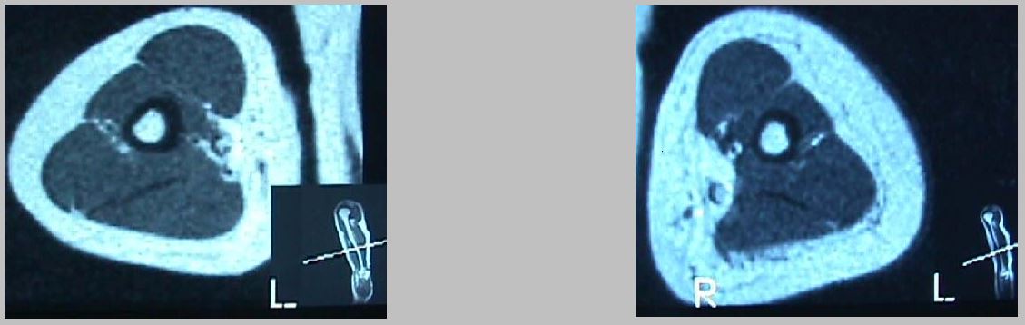
Cleavages in the adipose panniculus are observed on the involved side. A fibrous cutaneous tissue appeared in the extra-fascicular compartment.

Distally, closer to the elbow, the muscular-adipose tissue junction becomes more irregular.

Conclusion
we observed more adipose panniculus and fibrous tissue on the involved side. Furthermore, on the involved side adipose tissue invading the intra-fascicular compartment was also observed. Therefore, the search for an absolute oedema reduction with the conventional approach could be impractical. (cfr “Consensus document of the edema treatment”)“)